The “Mascara Sign”: Can Hounsfield Unit Help Diagnose Proximal Ventriculoperitoneal Shunt Malfunction in Paediatric Population?
DOI:
https://doi.org/10.5530/bems.9.1.2Keywords:
Shunt Malfunction, Shunt Revision Surgery, Hounsfield AttenuationAbstract
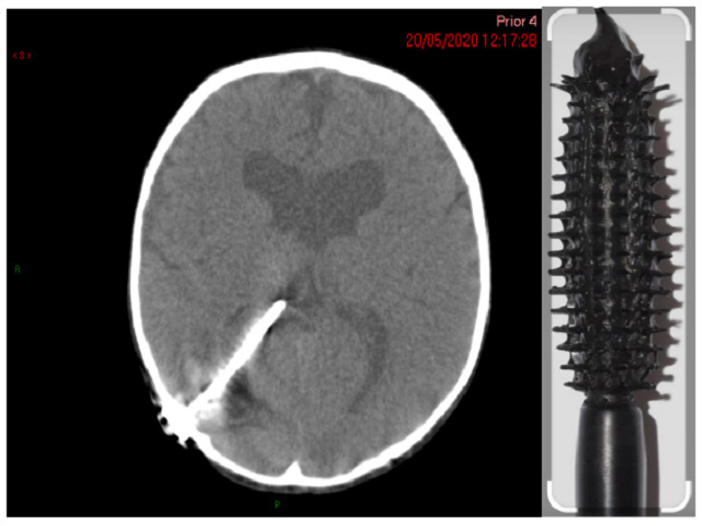
Background: Ventriculoperitoneal (VP) shunts are prone to malfunction, often requiring multiple shunt revisions. This is associated with significant morbidity, particularly in the paediatric population. This study aims to explore whether the appearance of a proximally blocked Ventriculoperitoneal (VP) shunt can be reproducibly established, using Hounsfield Unit of attenuation as a measure of density, from non-invasive CT imaging. The benefit of such a test would be to minimise the invasiveness of shunt surgeries on patients by localising the point of fault in a malfunctioning VP shunt. Materials and Methods: The data set of 174 paediatric neurosurgical patients with documented proximal VP shunt blockage was identified, with 16 patients meeting the inclusion criteria. CT. imaging was reviewed with an average density obtained for each of the 16 patients. A retrospective analysis was performed using the exact Sign test to compare the median difference in average intraluminal densities using Hounsfield attenuation during proximal catheter obstruction. Results: There was no statistically significant median increase in intraluminal density after a proximal catheter obstruction (p=1.00). In analyzing the CT scans, we have also observed that in some patients, there is a recognisable but subjective change in appearance of the proximal catheter when blocked, the “Mascara Sign.” Conclusion: Our retrospective pilot study demonstrated that Hounsfield attenuation cannot be used as an objective guide to identify proximal ventriculoperitoneal shunt obstruction on non-invasive CT imaging in the paediatric population.

Downloads
Published
How to Cite
Issue
Section
License
Copyright (c) 2023 Biology, Engineering, Medicine and Science Reports

This work is licensed under a Creative Commons Attribution-NonCommercial-NoDerivatives 4.0 International License.


